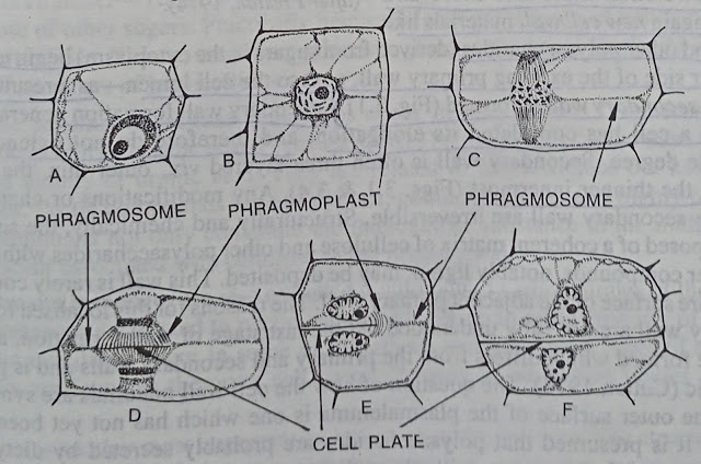
In highly organised plants, the cell wall is formed between the two daughter nuclei at the telophase stage of nuclear division by cytikinesis process.
 |
| Different stages in the formation of cell wall with successive layer |
According to Sharp (1943), Eames and MacDaniels (1947) and others origin of the cell wall is after the formation of daughter nuclei, protoplasmic matters of pectin in nature accumulate in the equatorial region in the form of droplet-like particles ; later on these particles increase in size and merge in course of time to form a continuous fluid plate, called cell plate.
According to Sinnott and Bloch (1941), during mitosis at the telophase stage, a fibrous structure known as phragmoplast appears between the two daughter nuclei which widens and becomes barrel-shaped.
At the same time, on the equatorial plane the cell plate begins to form inside the pharagmoplast. As soon as the cell plate enlarges the fibres of the phragmoplast approach the Wall of the dividing mother cell. Very soon the cell plate reaches the side walls of the mother cell but the contact with the end walls of the cells is delayed. When the cell plate reaches all parts of existing wall of the dividing cell, the phragmoplast disappears completely.
 |
| Different stages in the origin of the cell wall through the formation of phragmoplast |
But work with electron microscope indicates that vesicles formed by the dictyosomes appearently fuse to form the cellplate and this process continues at both ends until the Cell plate reaches the existing cell wall (Whaley and Mollenhauer, 1963 ; Muhlethaler, 1965). Elements of the endoplasmic reticulum become incorporated into the cell plate at regular intervals and indicate the positions of the future cytoplasmic connections (plasmodesmata) which traverse the wall between adjacent cell.
This cell plate, due to some physical and chemical changes is later converted into the middle lamella of the wall. Middle lamella thus forms the first partition wall between the two daughter cells arising through cell division and acts as an intercellular cement connecting the cells together.
Middle lamella is composed of pectic substances. Each of the protoplasts of the two cells now begins to secrete cell wall materials on or next to the middle lamella, as a result a soft and delicate wall known as primary wall is formed. The wall is usually single layered and chemically composed mainly of Celulose, hemicellulose and other polysaccharides. Primary wall grows both in surface as well as in thickness. The primary wall is not uniform in thickness throughout its extent but, on the other hand, shows thin areas of various patterns. At the time of the growth of the cell, the primary wall gets stretched more and more and ultimately again new cell wall materials like cellulose and other polysaccharides (derived from sugars in the cytoplasm) begin to deposit on the inner side of the existing primary wall next to the cell lumen-as a result another wall called secondary wall is formed. Secondary wall formation generally takes place after a cell has completed its elongation, and therefore do not extend to any considerable degree. Secondary wall is also three layered viz., outer thin, the thickest middle and the thinner innermost.
Any modifications or changes that occur in the secondary wa are irreversible. Structurally and chemically, the secondary wall is composed of a coherent matrix of cellulose and other polysaccharides within which various other compounds, notably Iignin, may be deposited. This wall is rarely continuous over the entire surface of the adjacent primary wall. The reasons for this localised formation of secondary wall are not fully understood. At the last stage of wall formation, a tertiary wall may be formed which differs from the primary and secondary walls and is probably not cellulosic (Cutter, 1978). The question of how the cell wall materials are synthesized and reach the outer surface of the plasmalemma is one which has not yet been clearly understood. It is presumed that polysaccharides are probably secreted by dictyosome-cisternae and move thence to fuse with plasmalemma ; vesicles of endoplasmic reticulum may also take part in this process (Northcote, 1972).
 |
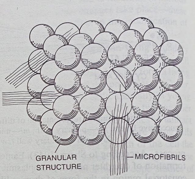
Arrangement of granular structure
|
According to recent view, small, ordered granules (comprising synthetases or cellulose-synthesizing enzymes) occurring on the outer surface of the plasmalemma are involved in the cellulose synthesis and orientation of microfibrils. When the glucan synthetases resent in the granules come in contact with the end of a microfibril, it is then extended terminally by condensation of glucose residues. On the basis of this concept, the orientation of microfibrils is determined by square packing of the granules. Precursors of cellulose are derived from dictyosome vesicles, endoplasmic reticulum or microtubules (Preston, 1974).
The occurrence of such granules has been observed in xylem, collenchyma and parenchyma cells of higher plants.
Now-a-days, cell wall formation has also studied in isolated plant protoplasts grown in culture media. In such isolated protoplasts, cell wall formation in initial stage is not associated with cytokinesis, here cell plate is not formed but the wall is formed at the surface of the plasmalemma. At first a membrane is formed, probably due to the action of the dictyosomes ; next cellulose microfibrils are formed between the outer envelope and the plasmalemma (Cocking, 1974).


Comments
Post a Comment