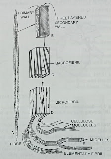
The principal component of cell wall is cellulose, the ultra structure of cell walls is therefore based on cellulose. Work with electron microscope shows that cellulose in cell walls occurs in the form of long-chain molecules (Roelofsen, 1959; Albersheim, 1975; Frey-Wyssling, 1969 -76; Frey-Wyssling and Muhlethaler, 1965). These chain like molecules may be arranged randomly or in a more or less regular fashion. Each such cellulose molecule has 8Å maximum width. Again cellulose molecules are regularly arranged in bundles-each such bundle forms an elementary fibril. Each bundle of elementary fibril contains 40-100 cellulose molecules in a transection, and is about 3.5 nm wide and 3 nm thick-some authors suggest that it has a widest diameter of 100Å.
Both the cellulose molecules and the elementary fibrils are ribbon-like structures. Within the elementary fibrils themselves are smaller units termed micelles or crystallites (Wardrop, 1962), which are small aggregations of cellulose molecules that lie parallel to one another and thus confer a crystalline structure upon the elementary fibril ; only very small part of elementary fibril, that are presumably arranged at random, may be paracrystalline. It was observed that the number of glucose residues in cellulose molecules of fibre cell varies from 500 to 10,000 and the length of such molecules varies from 0.25-57um.
 |
| Ultrastructure of the cell wall |
The elementary fibrils are again arranged in bundles, each such bundle is called microfibril which is 250Å wide and contains 2,000 cellulose molecules in transection. Microfibrils are combined into macrofibrils, each of which is 0.4u wide and contains 500,000 cellulose molecules in transection. In case of the secondary wall of ramie (Boehmeria) fibre, about 2000,000,000 cellulose molecules were found in a transection. This is the structure that has been studied in case of the secondary wall.
The primary wall is similar in structure to the secondary wall in that it consists of crystalline (anisotropic) cellulose microfibrils and a noncellulotic matrix.
Microfibrils consists of bundles of cellulose molecules partly arranged into orderly three dimensional lattices i.e. the micelles. The crystalline properties of micelles are due to regular spacing of glucose residues which are connected by b-1-4 glucosidic bonds.
The spaces between the randomly arranged molecules in the microfibril are filled up with water, pectic substances, hemicellulose and in secondary walls lignin, cutin, suberin etc. The swelling of the cell wall during lignification is mainly due to this depositions of lignin between the existing cellulose framework of the wall. The occurrence of a group of proteins containing hydroxyproline in the primary walls of various tissues has also been demonstrated by N. J. King and S. T. Bavley (I965), these proteins may serve enzymatic as well as structural functions. The protein may be involved in the orientation of the fibrils (Muhlethaler, 1967). Many workers think that the walI-protein plays an important role in cell extension, and accordingly that protein has given the name “extensin”. Experiments with radioactive isotopes suggests that the protein is deposited throughout the wall matrix (Roberts and Northcote, 1972). In most dicotyledons the cellulose microfibrils are coated with a single layer of hemicellulose of the nature xyloglucan. The hemicellulosc is cross linked by pectic polymers, so that elementary fibrils are interconnected. In monocotyledons similar structure are found except that arabmoxylan takes the place of xyloglucan (Albersheim, 1974).
 |
Arrangement of microfibrils
|
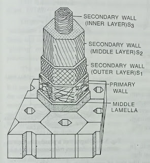
The microfibrils are arranged variously in cell walls. In the secondary walls they are arranged more regularly. In the primary walls microfibrils are often arranged in a direction more or less transverse to the longitudinal axis. The microfibrils become arranged more longitudinally durin g growth of the cell. Microfbrils become oriented more and more longitudinally when even subsequent wall layers are formed. This transition is gradual.
Studies of cell walls of different cell types stained with permanganate have shown that in parenchyma cell walls the fibrils are laid down in lamellae-here the direction of fibrils alternate in successive lamellae ; walls of sieve elements and collenchyma cells are polylamellate (Deshpande; 1976).
Different orientation of the wall layers in the secondary wall is also observed ; here the layer S1 is the outermost and adjacent to the primary wall ; within this layer S2 and S3 are laid down-in S1 and S3, the fibrils forms a lax helix while in S2 fibrils form a steep helical structure.


Comments
Post a Comment