The thickening of the cell wall takes place due to the deposition of secondary cell wall materials like cellulose, lignin, cutin, suberin, etc over the primary cell wall and finally the cell attains its full size and the wall becomes thick. The depostion of the secondary cell wall materials is localised in many cases, specially in case of cells and vessels of xylem tissue-here the secondary cell wall materials are not uniformly deposited on the primary cell wall, rather they are localised to certain regions on the primary cell wall.
Thereby some portion of the cell wall remains thin and other parts remain thick. As a result, various patterns and designs are formed due to such localised thickening of the cell wall. Thickening gives strength to the cell wall and can withstand internal pressure as well.
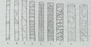
Various patterns of markings are usually found on the inner surface of the cell wall, such as :
(a) annular, (b) spiral, (c) scalariform, (d) reticulate and (e) pitted.
 |
| Various types of thickening of the cell wall in longitudinal sections. |
In annular or ring-like type of thickening, the secondary thickening matters are disposed centripetally in the protoxylem elements in the form of rings at regular intervals, e.g. annular vessels of wood or xylem.
In spiral thickening, the thickenings (due to the deposition of secondary thickening matters) take place inside the wall in the form of spiral bands-this type is also found in protoxylem elements.
In the scalariform type, secondary thickening matters are disposed like the steps of a ladder, both in longitudinal and transverse directions.
Reticulate type of thickening results from irregular deposition of secondary thickening substances inthe form of a network.
Pitted-In this type small, thin and unthickened areas, called pits, are formed on the primary wall due to the deposition of the secondary wall materials all over the primary wall except within some areas which become thin and remain unthickened.
OTHER TYPES OF WALL THlCKENlNG
(a) Trabeculae-These are rod-like or bar-like projected thickening of the wall which traverse the cell lumen radially. Trabeculae generally occur in radial rows in the wood elements.
They are frequently present in the tracheids of the secondary wood of conifers.
 |
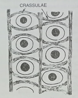
| Crassulae in tracheids of pinus sp. |
(b) Crassulae-These are linear or crescent-shaped thickenings of middle lamella and Primary wall which occur along the outer and lower margins of pit-pairs. Crassulae may encircle the pits some-times. They represent the borders of the primary pit-fields of the young cells from which the element developed.
Previously crassulae were designated as “bars of Sanio ” and “rims of Sanio” after the name of the anatomist Sanio, But these terms are now confusing in usage as these terms are also applied to trabeculae. Crassulae are found in the tracheids of some gymnosperms.
(c) Wart structures-These are wart-like structures that have been observed on the inner surface of the secondary wall of tracheids of certain conifers and of fibres and vessels of many dicots (Liese and Ledbetter, 1963 ;Wardrop et al, 1969). Wart structures develop after or towards the end of the differentiation and lignification of the secondary wall. These structures vary between 0.1u and 0.5u in diameter. According to some authors they consist of remnants of the protoplast.


Comments
Post a Comment